APPLICATIONS
PRISM Applications
Discover PRISM unique capabilities and fields of application.
Image Scanning microscopy (ISM) and Fluorescence Lifetime Imaging (FLIM) provided by PRISM have a wide range of applications in science. They are crucial for cellular and molecular biology, where super-resolution microscopy combined with fluorescence lifetime imaging can be used to study the structure and dynamics of biological molecules and cellular processes. They play a major role in neurobiology, where they are used to image brain tissue and neural processes. They find application in materials science to study the properties of various materials at the nanoscale level. Among others, they also represent the optimal imaging choice for nanotechnology, biophysics, pharmacology, plant science, and more. Continue reading to discover all PRISM application fields.
APPLICATION #1
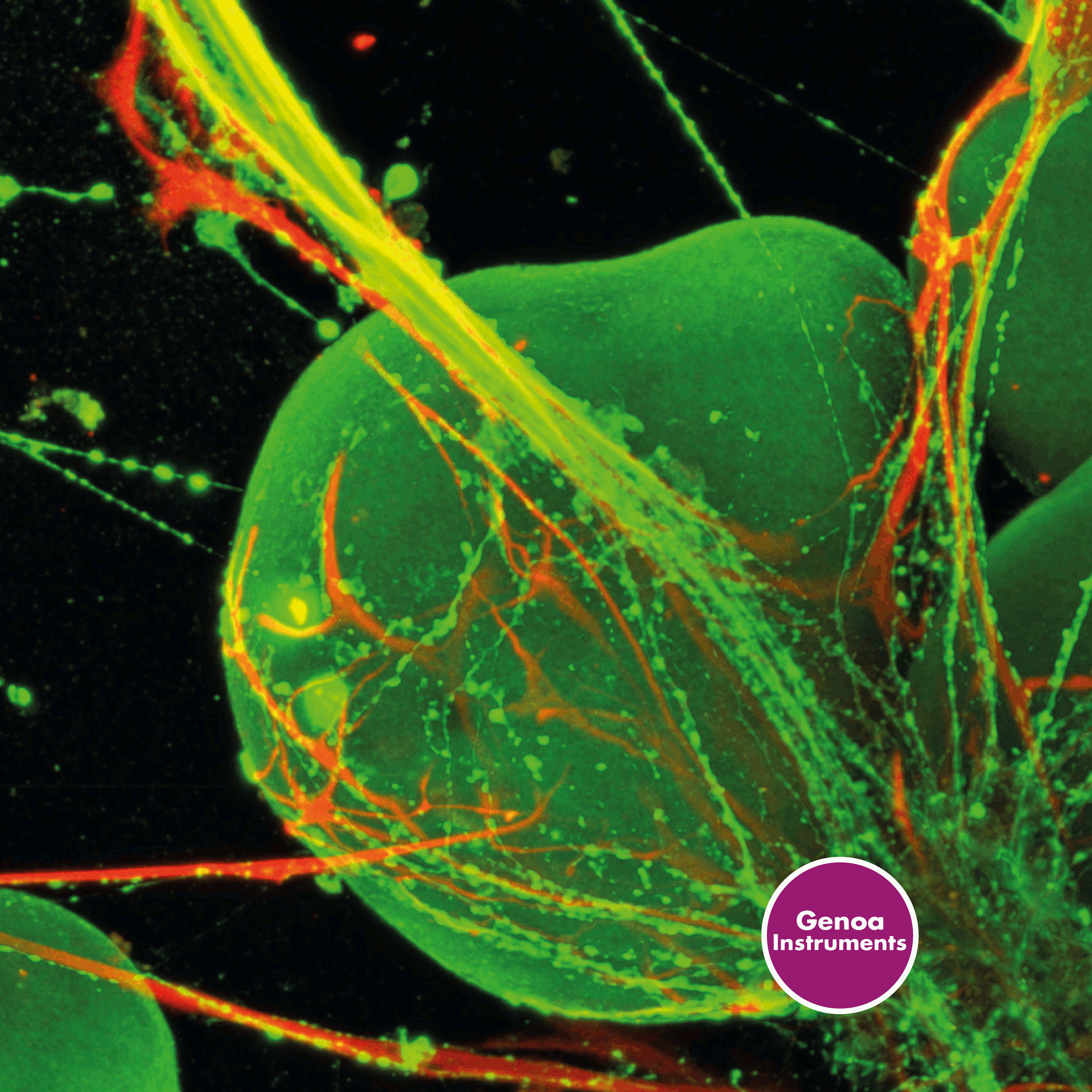
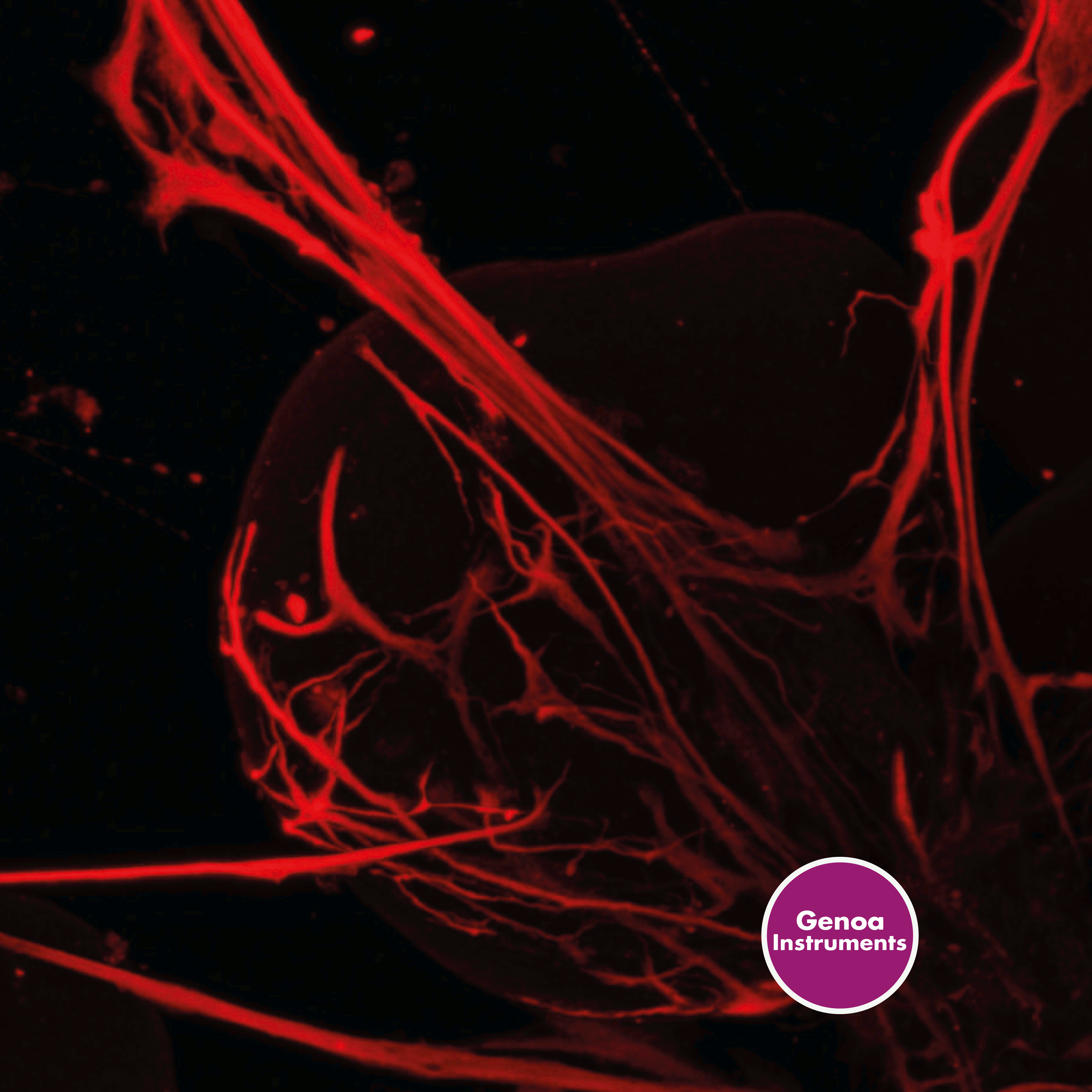
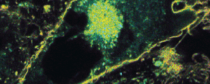
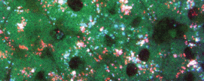
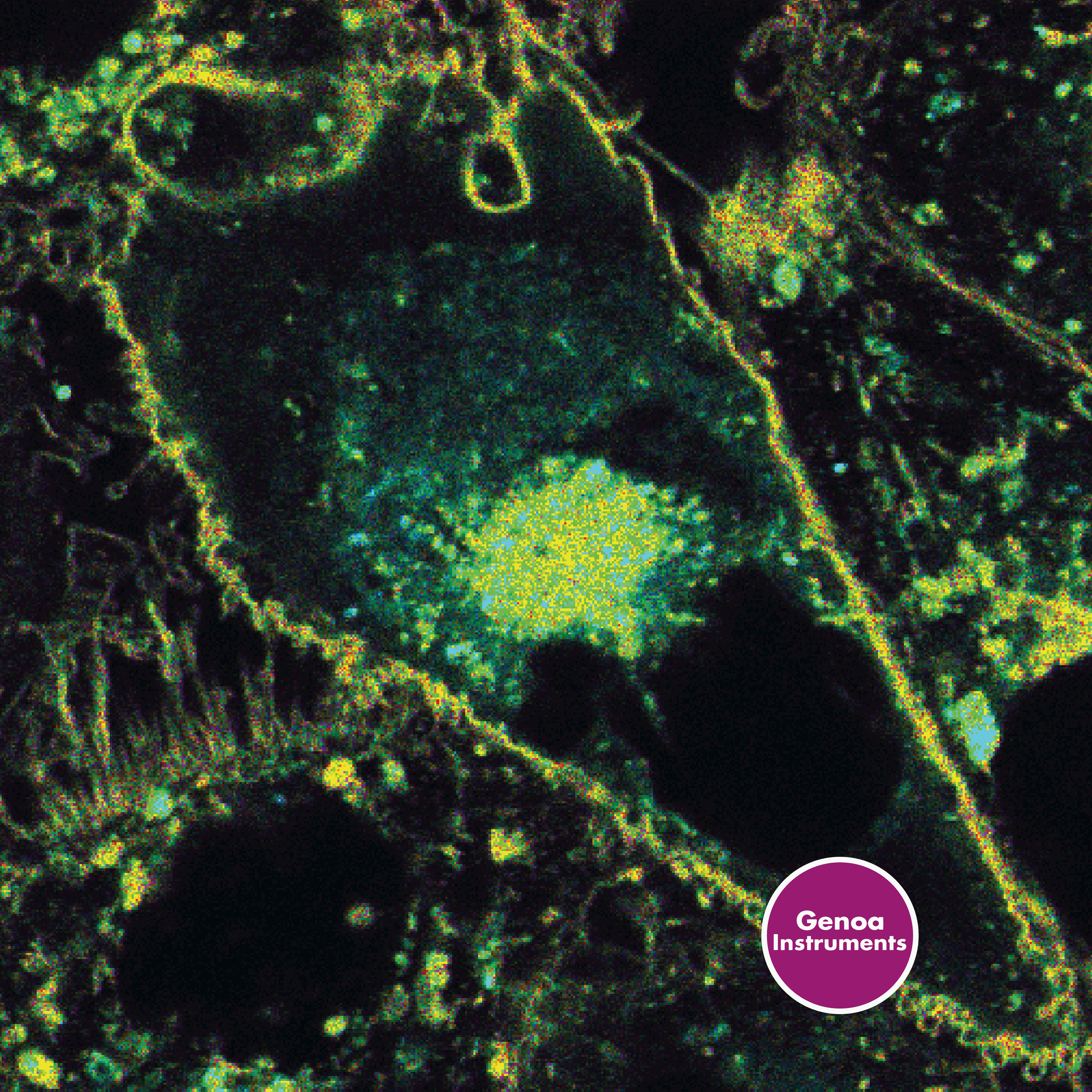
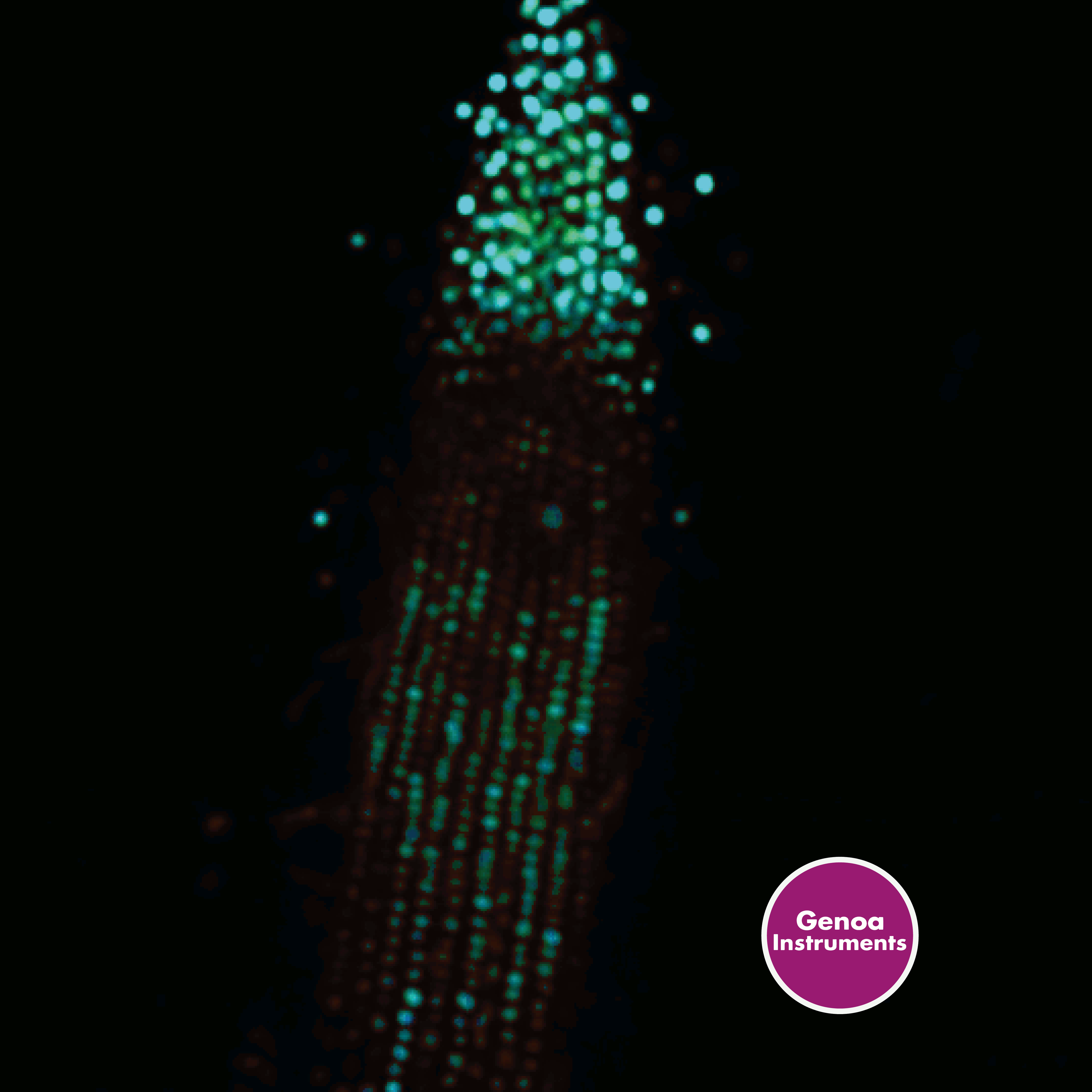
Cell Biology
Time-resolved super-resolution at your service.
Super-resolution optical microscopy has revolutionized the field of cellular and molecular biology enabling scientists to study structures and processes at the nanoscale level. With conventional light microscopy, the resolution is limited by the diffraction of light, which results in a maximum resolution of about 200 nanometers. Super-resolution optical microscopy techniques, such as Image Scanning Microscopy, break this resolution barrier offering a resolution up to ten times higher than conventional microscopy. This improved resolution allows researchers to investigate cellular structures and processes that were previously beyond the reach of conventional microscopy. With PRISM, you can double the resolution of a traditional confocal microscope and reach a spatial resolution of 100 nm, expanding research opportunities and advancing our understanding of fundamental biological processes.

APPLICATION #2
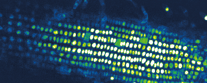
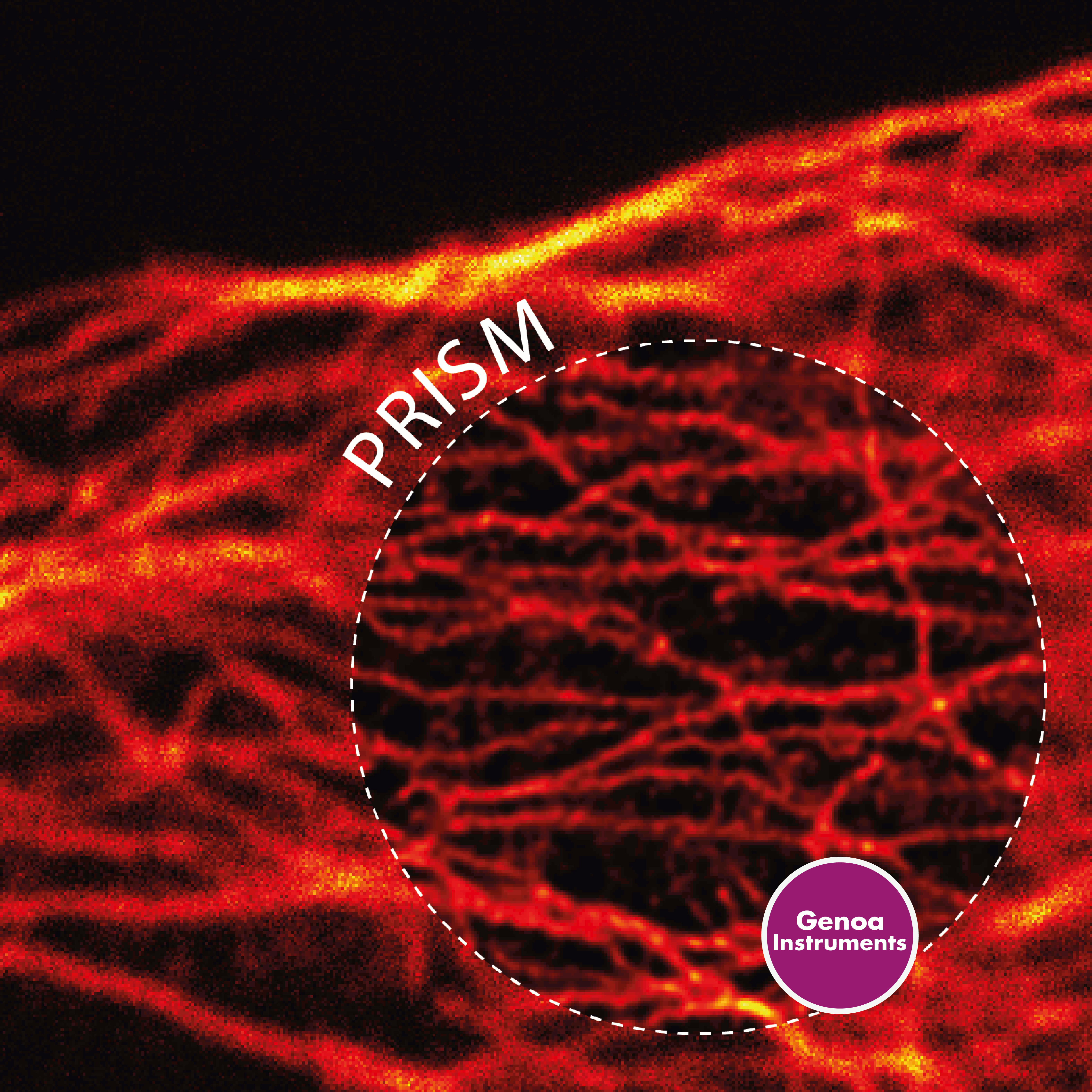
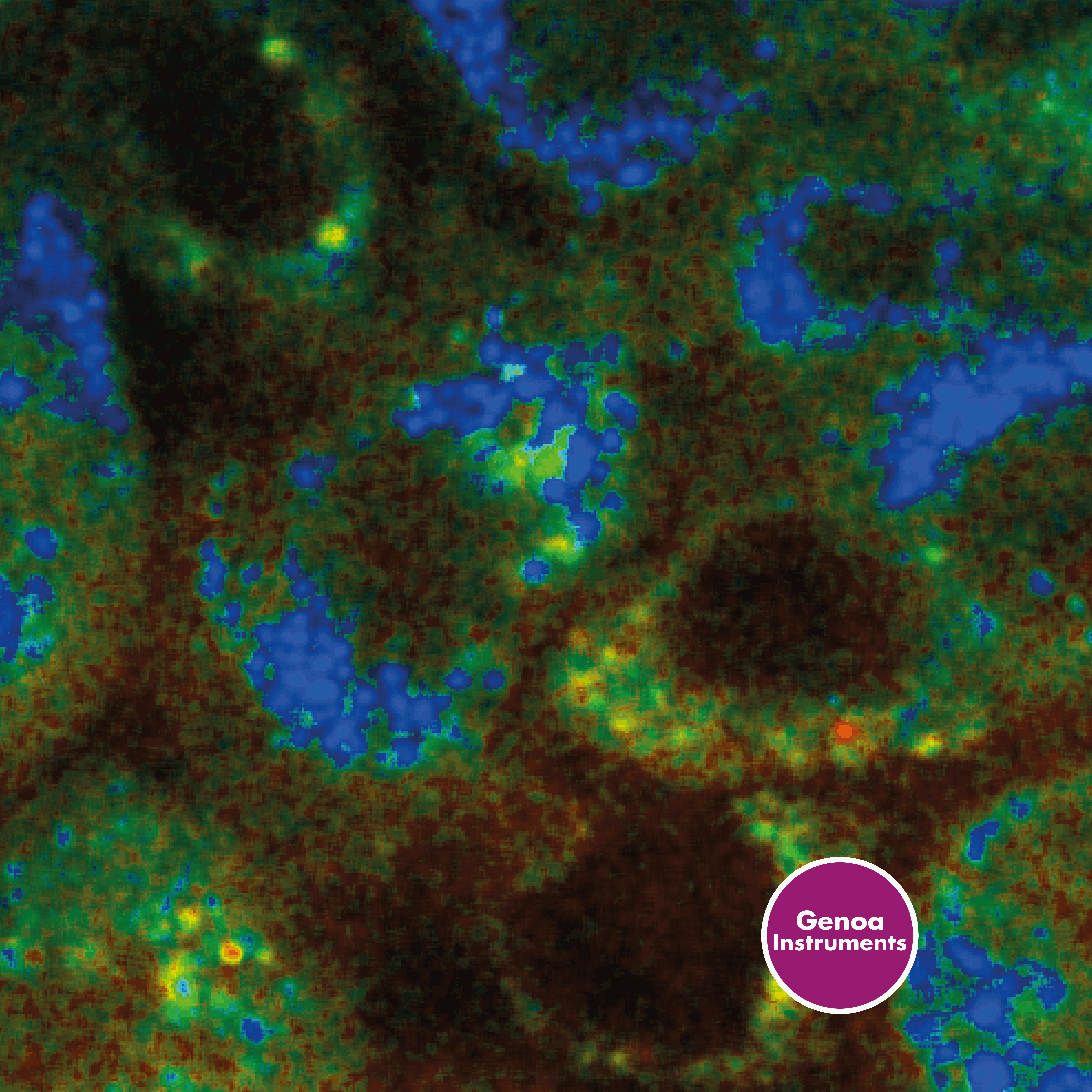
Lifetime Imaging
Natural lifetime experiments.
Fluorescence lifetime imaging (FLIM) is of utmost relevance for cellular and molecular biology as it provides quantitative information about the fluorescence properties of biological molecules. FLIM measures the fluorescence lifetime of molecules - the amount of time a molecule spends in an excited state following light stimulation. This measurement can be used to identify a wide variety of molecules and biomarkers, monitor changes in molecular conformation or concentration, and study molecular interactions. FLIM is a precious tool for studying biological processes in real-time, making it extremely useful for live-cell imaging. By combining FLIM with super-resolution via Fluorescence Lifetime Image Scanning Microscopy - FLISM, PRISM provides unprecedented quantitative information on the structures and dynamics of biological molecules.

APPLICATION #3
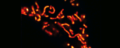
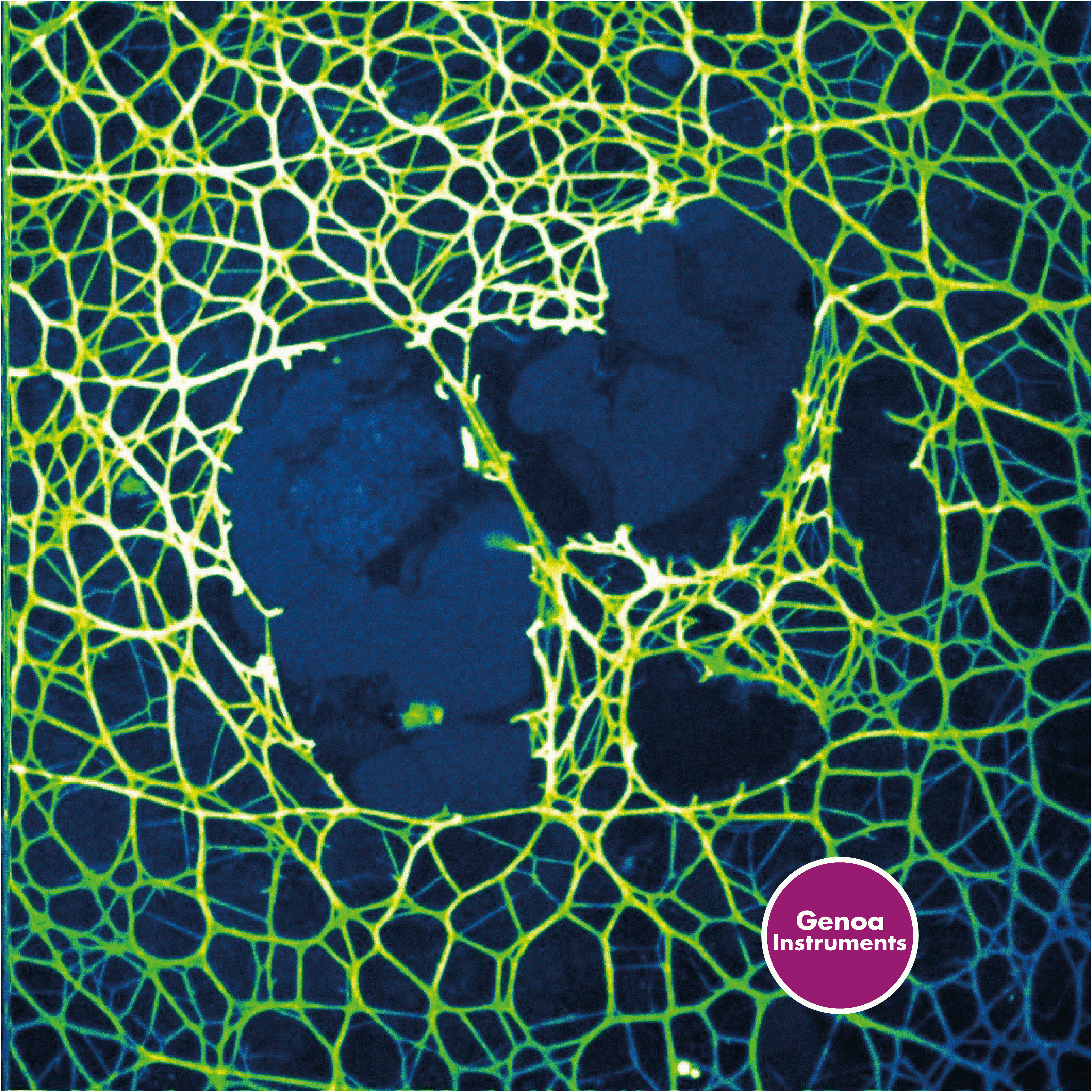
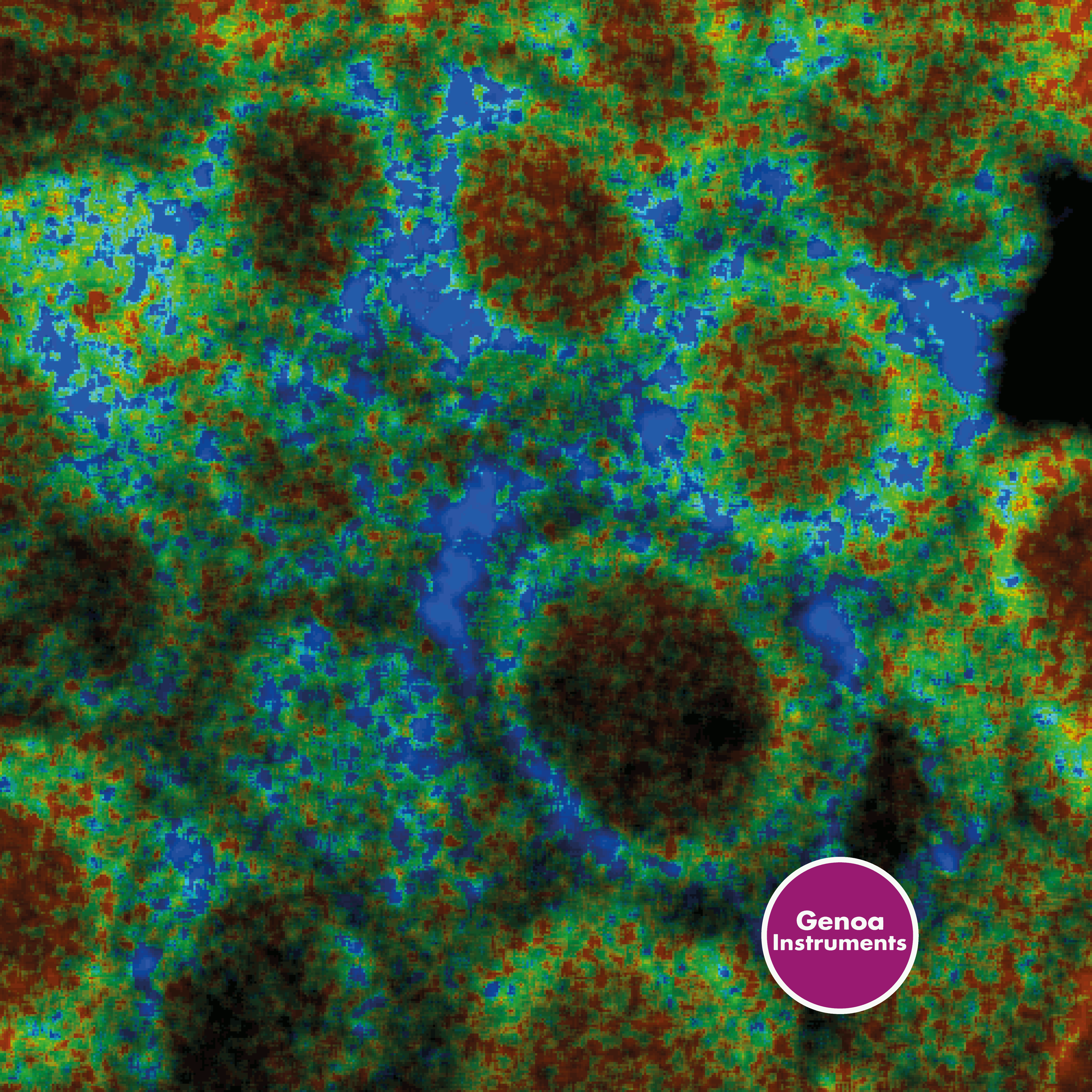
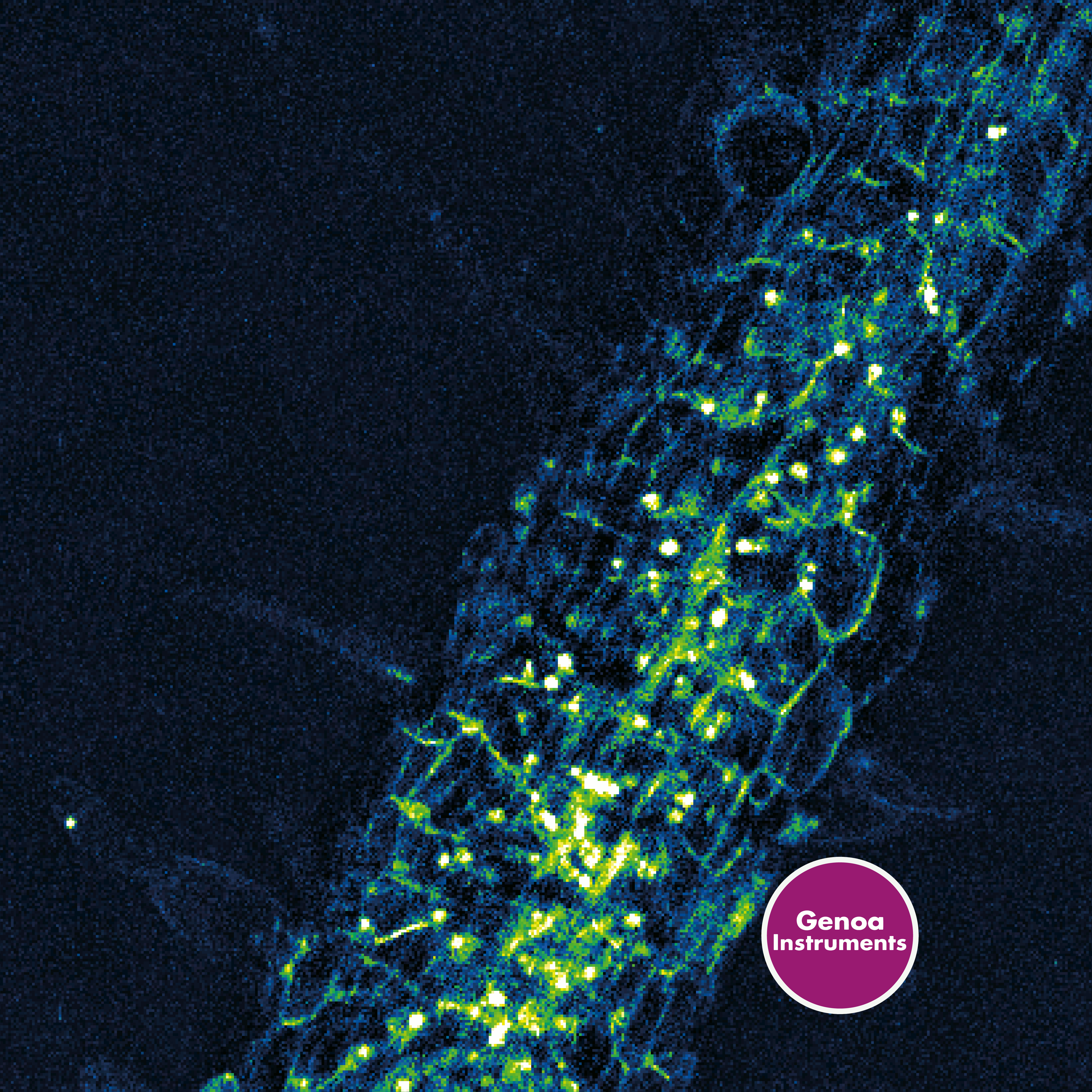
Live-cell imaging
Be gentle on your sample, with great contrast.
With less photodamage and photobleaching compared to other super-resolution techniques, Image Scanning Microscopy finds natural application in live-cell imaging. ISM is gentler on the sample and provides high-contrast images thanks to its optimal signal-to-noise ratio in case of dim samples (no pinhole). PRISM enables achieving the same resolution of a confocal closed-pinhole image while using one-tenth the illumination intensity. This allows to observe dynamic processes of living cells with high spatial and temporal resolution, and can provide critical insights into biological processes such as protein dynamics, organelle interactions, and signaling pathways. The gentle treatment of samples allows for longer imaging times and minimizes the impact of imaging on cell physiology. In addition, the high-contrast images obtained through ISM offer excellent structural information, enabling the identification of previously unseen structures and the mapping of cellular components at a nanometer scale. PRISM allows for super-resolution time-lapse imaging in live cells with negligible photobleaching (<5% after ten frames) while traditional confocal imaging could achieve a similar resolution only by using 10 times greater illumination power, leading to strong photobleaching (>75% after ten frames) and phototoxicity. ISM is a powerful tool that has revolutionized our ability to observe and understand the intricacies of live-cell biology.
Time-lapse imaging of mitochondria
signal-to-noise
resolution
signal-to-noise
resolution
signal-to-noise
resolution
APPLICATION #4
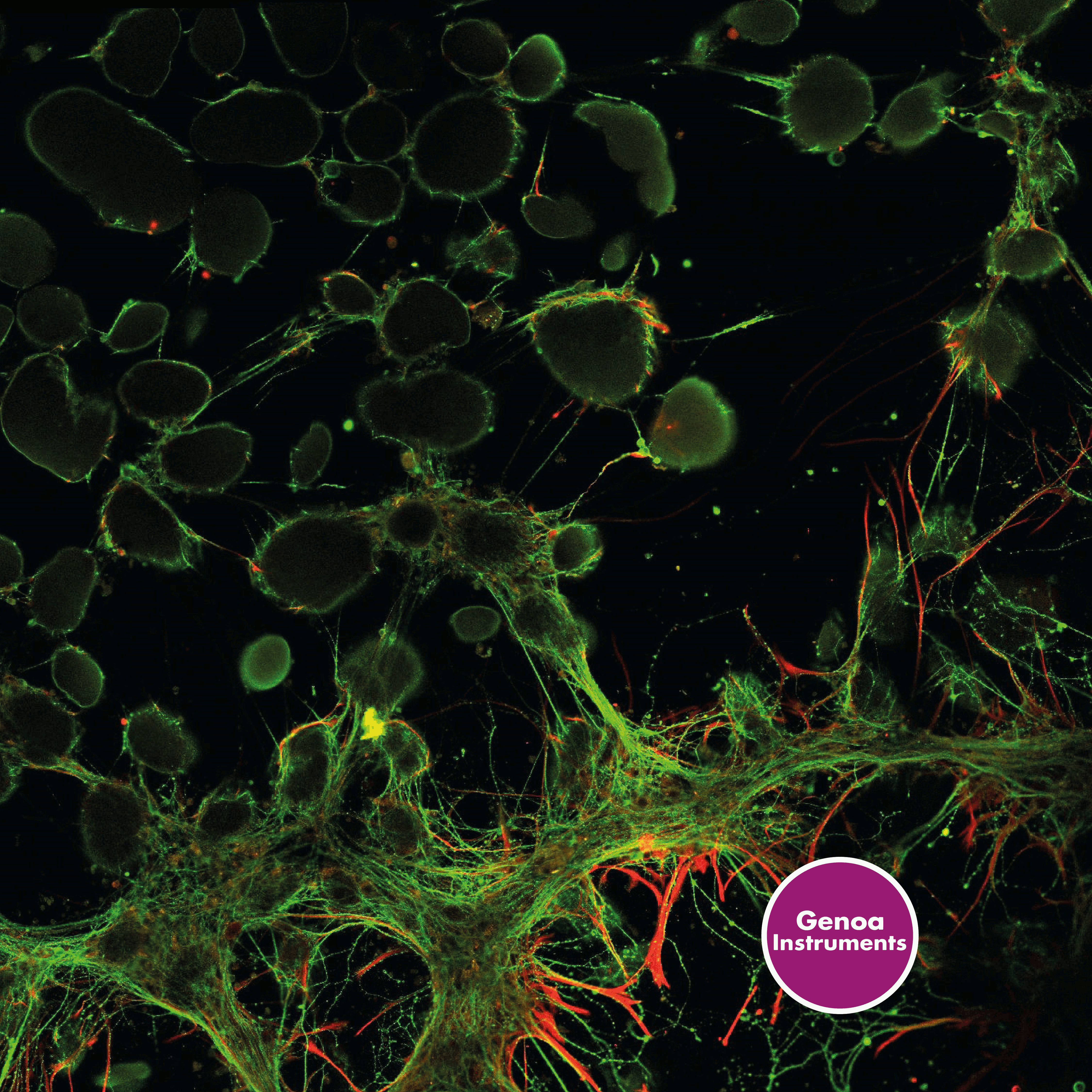
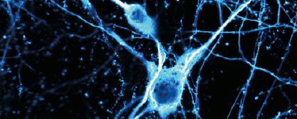
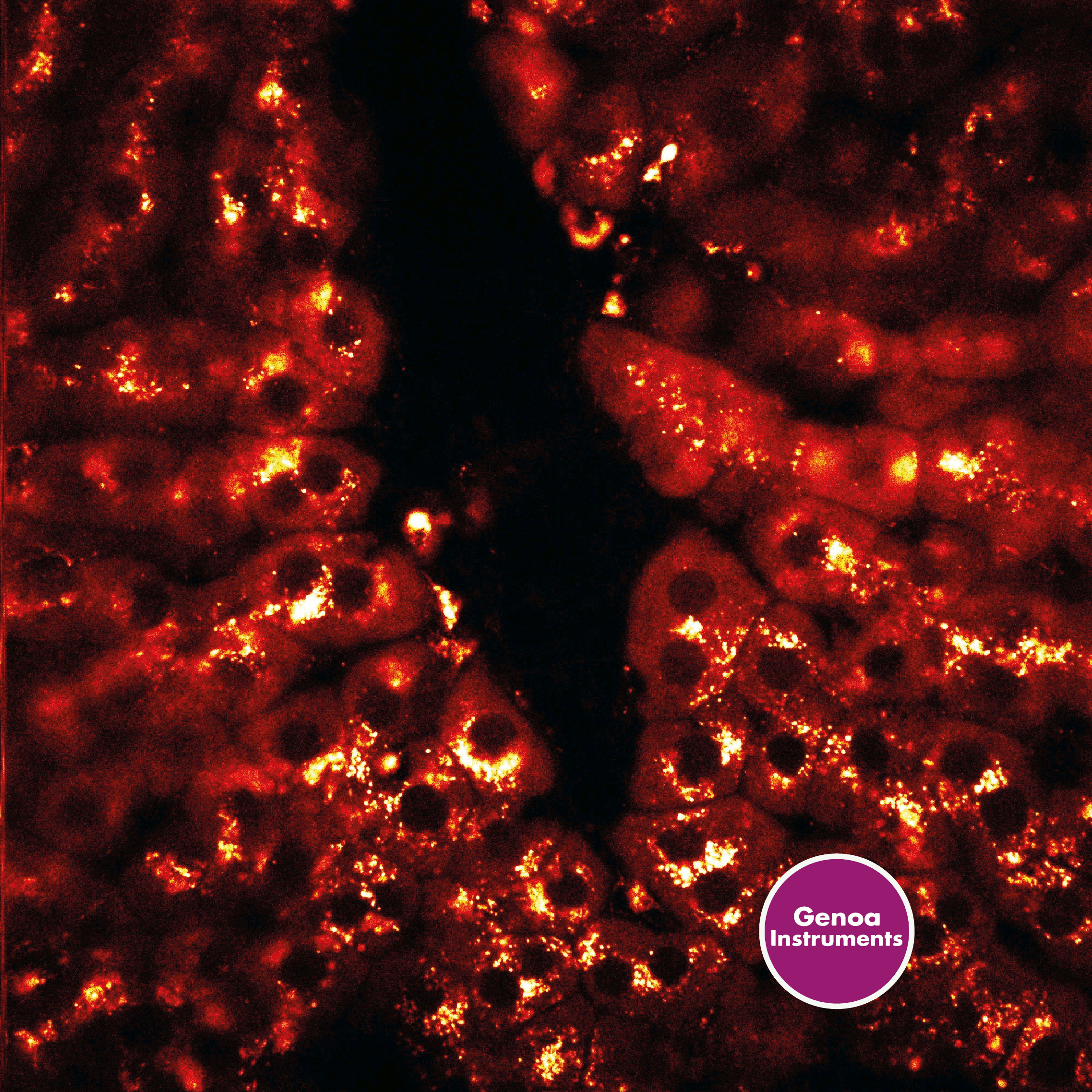
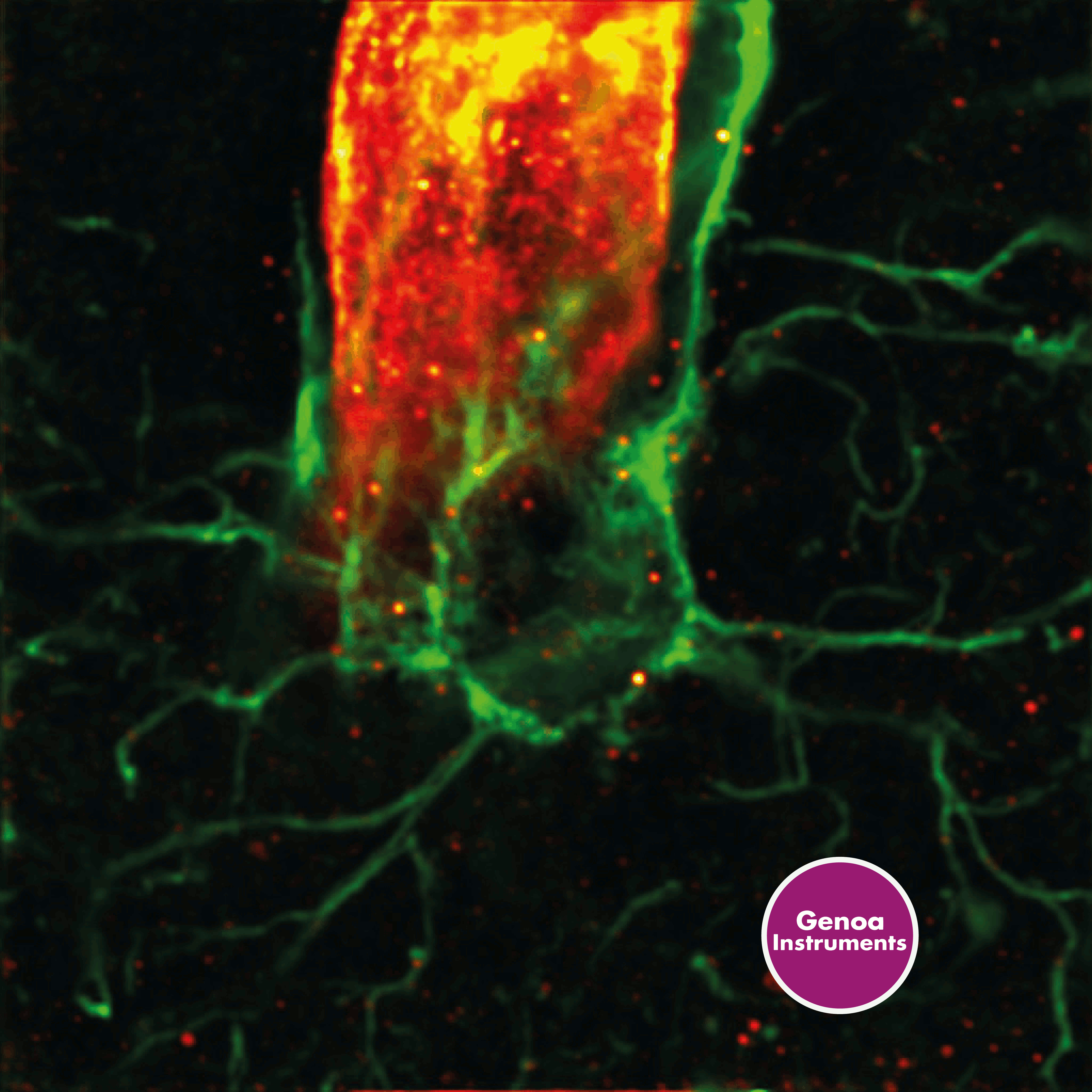
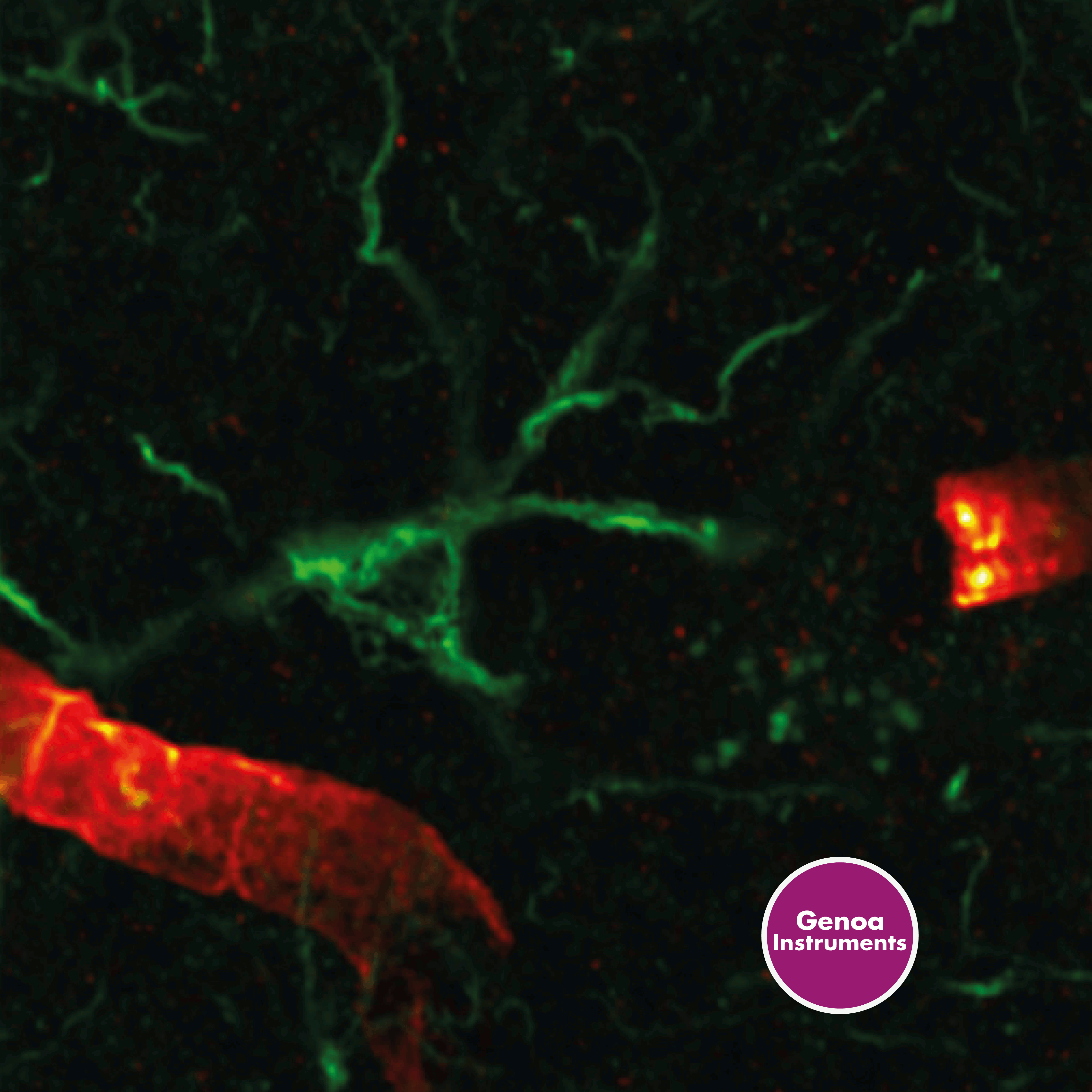
Neurobiology
Deep imaging at its best with ISM.
Study the structure and function of neurons and the nervous system. PRISM can be your perfect companion when performing deep imaging of neuronal processes in brain tissues, allowing to resolve neuronal structures which typically fall below the resolution limit of conventional light microscopes. Image Scanning Microscopy can provide terrific signal-to-noise ratio enhancement, enabling a 2-fold resolution improvement also in scattering samples and low-light conditions. for these reasons, ISM is emerging as a great technique in neurobiology due to its ability to provide high-resolution images with a high signal-to-noise ratio. Compared to other super-resolution techniques, ISM stands out when imaging deep within scattering tissues, such as the brain, due to its ability to reject out-of-focus light. Once again, ISM is gentle on samples and induces less photodamage and photobleaching, making it ideal also for live-cell imaging of neuronal activity. These features have made ISM a popular tool in the field of neurobiology, enabling researchers to study the complex dynamics of neuronal networks with unprecedented spatial and temporal resolution.


APPLICATION #5
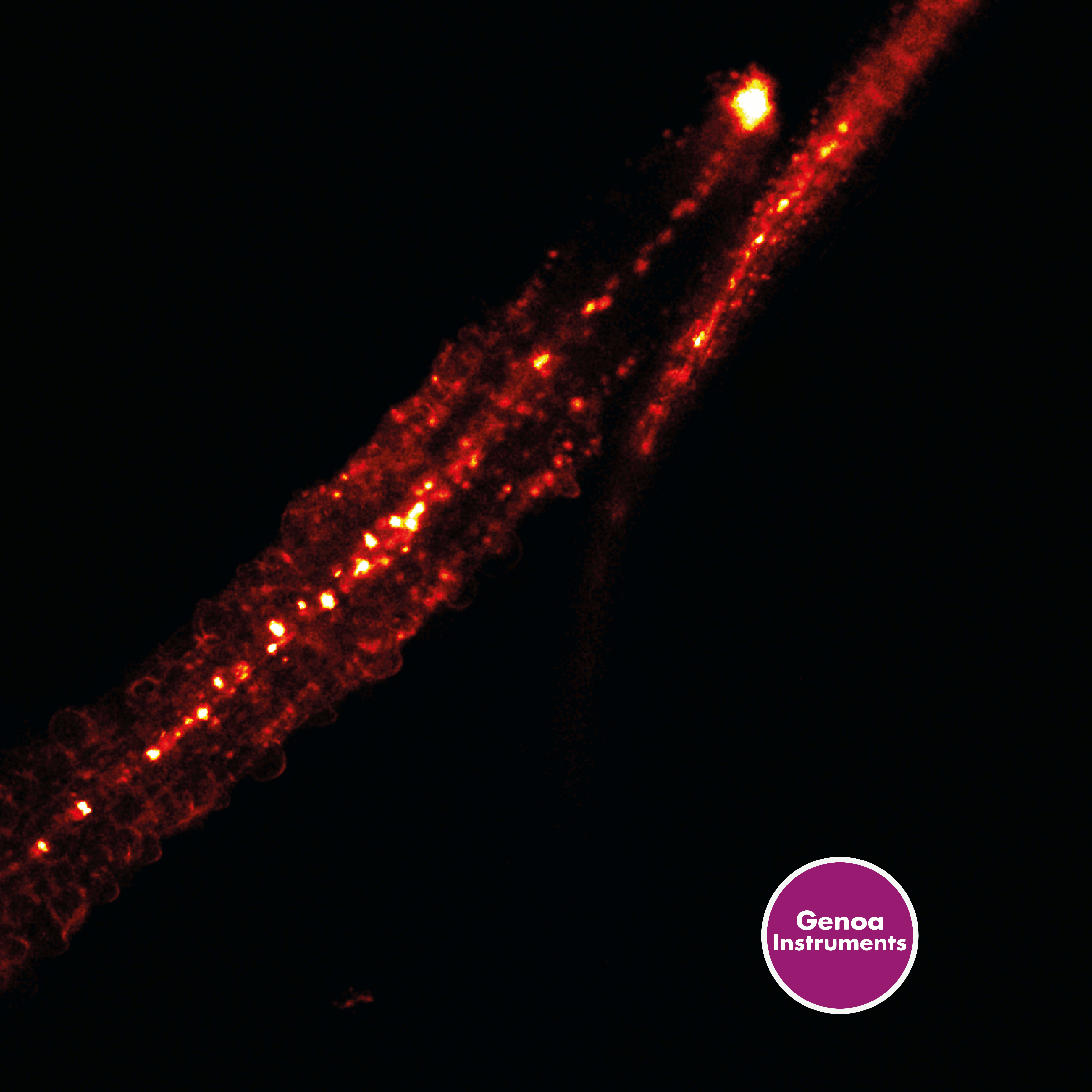
Plant Biology
Different scales, excellent performance.
Use PRISM imaging capabilities to investigate plant biology across a wide range of scales. Super-resolution optical microscopy is becoming more and more popular in plant science as it provides the ability to observe morphological structures of plants with unprecedented detail. For example, it allows for the visualization of nanoscale cellular structures such as chloroplasts and mitochondria, and can also be used to study the dynamics of cell wall organization and protein localization. Additionally, when combined with lifetime measurements as in PRISM, fluorescence lifetime imaging (FLIM) can further improve studies in the field by enabling investigation of the intrinsic autofluorescence signal of numerous plant structures. This can provide quantitative information on important aspects of plant physiology, such as photosynthesis and plant stress responses. The super-resolution imaging and fluorescence lifetime imaging offered by PRISM are powerful tools for advancing our understanding of plant biology and can have important implications for crop improvement and environmental sustainability.

APPLICATION #6
Materials Science
Different scales, excellent performance.
Did you know that PRISM technology can be helpful for materials science? Super-resolution imaging combined with time-resolved measurements can be particularly valuable in nanotechnology and biomaterials. For example, super-resolution can be used to study the structure and behavior of individual nanoparticles, providing new insights into their properties and applications. Additionally, it can be used to study the interactions between materials and biological systems, such as the interactions between nanomaterials and cells or tissues. By providing high-resolution images, Image Scanning Microscopy can help to identify risks and opportunities for the development of new materials for biomedical and environmental applications. Another important application is the study of microplastics - small plastic particles - that are becoming an increasing environmental concern. Super-resolution microscopy combined with spectroscopic techniques such as FLISM (Fluorescence Lifetime Image Scanning Microscopy) by Genoa Instruments, can be used to study the structure and behavior of microplastics in a variety of environments, including water, soil, and biological systems. By providing detailed images of the size, shape, and distribution of microplastics, PRISM can help identify contamination sources and track microplastics movement through different environments, and develop new methods for removing microplastics from the environment. This can contribute to sustainability and planet-saving oriented research.