Image Scanning Microscopy (ISM) is an optical imaging technique that can provide super-resolution images of biological samples. It achieves high-resolution images by scanning a focused laser beam across the sample, while detecting the emitted light from the sample with a SPAD (Single Photon Avalanche Diode) array. The use of SPAD arrays allows for fast and sensitive detection of fluorescence signals, providing high-resolution images even with low-light samples. Additionally, ISM can naturally be combined with time-resolved measurement, such as fluorescence lifetime imaging (FLIM), to provide additional information on molecular interactions and dynamics within the sample. Compared to other super-resolution techniques, such as STED or SIM, ISM is gentle on the sample and induces less photodamage and photobleaching, allowing for longer imaging times and less damage to the sample. Furthermore, ISM can provide great contrast for imaging challenging samples, such as those with low fluorescence signals or those with high background fluorescence. Therefore, ISM is an advantageous imaging technique for live-cell imaging and a range of other applications in biology and material science.
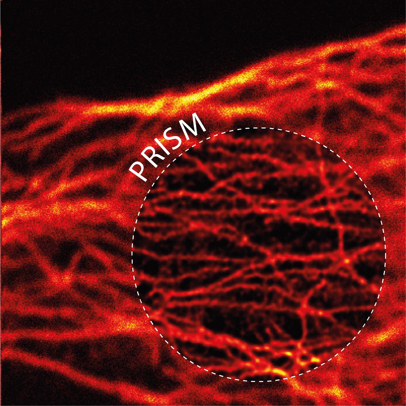
Image Scanning Microscopy (ISM) can improve the effective spatial resolution of conventional confocal laser scanning microscopy to its theoretical limit, without compromising any of its functionalities.
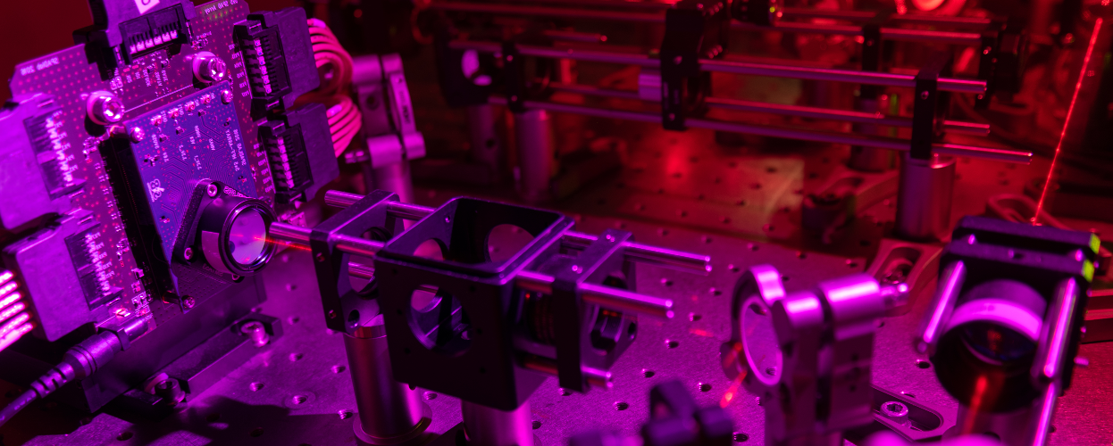
Our team implemented a natural design of ISM based on a fast single-photon detector array which allows straightforward upgrade of confocal microscopy and provides access to the raw scanned images, also paving the way to adaptive reconstruction methods.
While standard all-optical implementations of ISM can reduce versatility, our solution enables for a direct combination with fluorescence spectroscopy techniques, such as fluorescence lifetime imaging and fluorescence correlation spectroscopy.
Push confocal microscopy resolution to its theoretical limit by achieving a 2-fold improvement.
Achieve the same resolution as ideal confocal at one-tenth the illumination intensity.
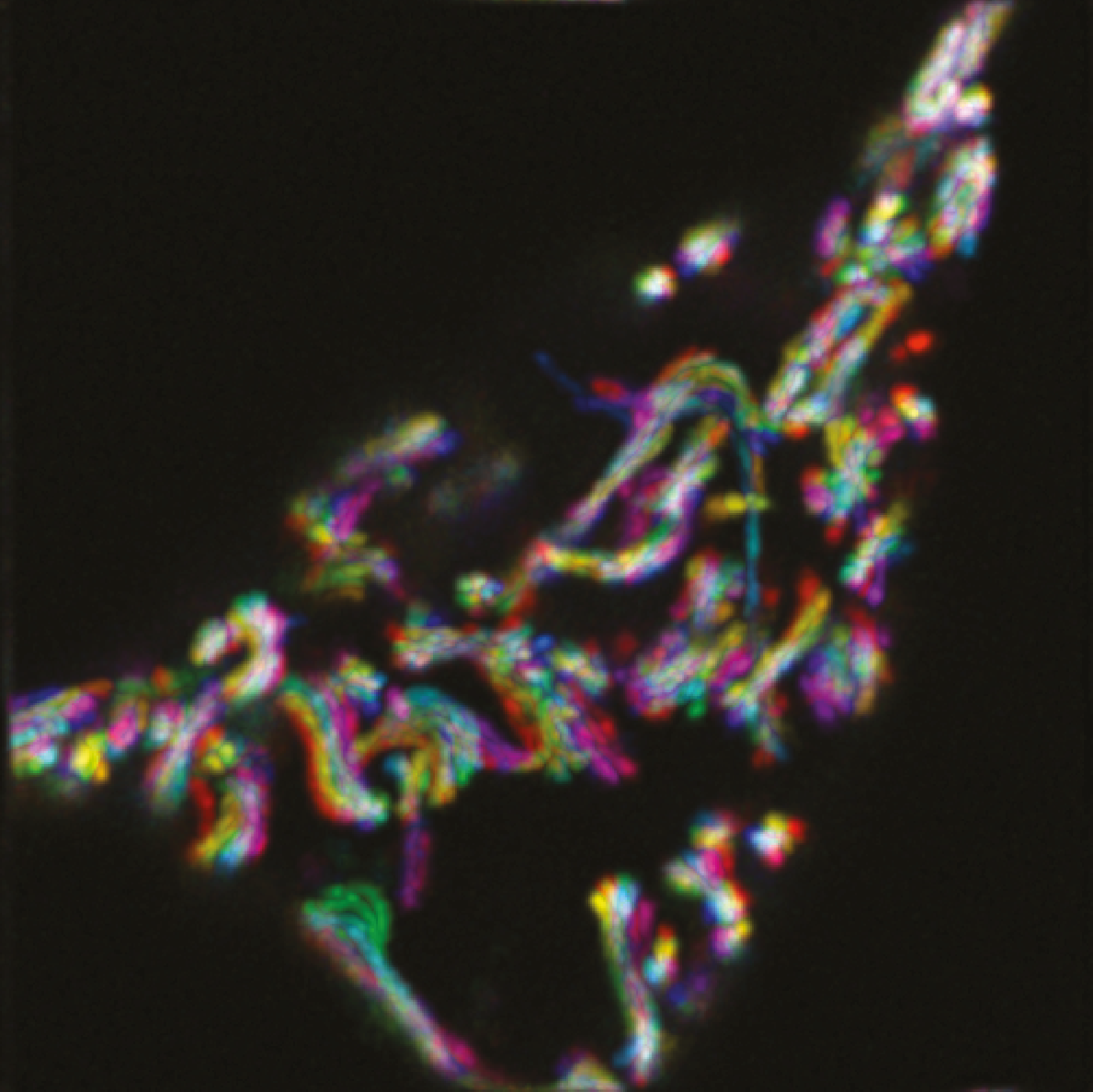
Perform time-resolved imaging while maintaining spatial super-resolution.